FTIR Spectroscopy - General Description
Fourier Transformed Infrared (FTIR) Spectroscopy is an optical technique used to study the electrodynamic properties of solids. FTIR Spectroscopy allows investigating the response function of a material, i.e. optical conductivity, as a function of frequency. For this reflectance or transmittance spectra are analyzed and interpreted for their physical meaning. Flexible integration, good SNR, a wide spectral range, and high resolution make FTIR spectroscopy a powerful experimental method in modern condensed matter physics.
Principle
A typical FTIR setup contains a broadband light source, detector, and an interferometer (for instance, Michelson type). By employing different types of sources and detectors, the reflectance or transmittance of the sample can be measured over a broad spectral range from around 20 cm-1 to over 25000 cm-1.
A broadband light beam is guided into the interferometer, consisting of a fixed mirror, a movable mirror, and a beam splitter. Depending on the position of the movable mirror (scanner), wave interference modulates the recombined beam, resulting in an interferogram (intensity over path difference). For a precise measurement of the optical path difference between the interferometer arms, a HeNe laser is used. If a sample is placed in the beam path, the interferogram is modified due to the optical properties of the sample.
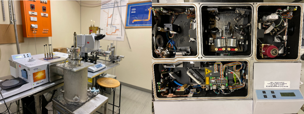
IFS 113v
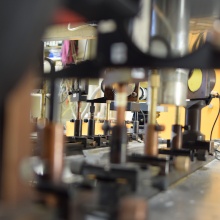
The IFS 113v is used for recording high-resolution spectra in the FIR range from 20 cm-1 to 1000 cm-1. Multiple cryogenic systems are combinable with the spectrometer. For instance, a LiHe-flow cryostat equipped with an in-situ gold evaporation unit allows to precisely measure the reflectance of a sample down to temperatures as low as 3 K. For transmission geometry, a LiHe-bath cryostat is used. As well, the spectrometer can be operated together with a superconducting magnet (Oxford instruments) and LiHe-flow cryostat to measure reflectance or transmittance under external magnetic field (up to 7.5 T) at low temperatures.
Fourier Transformation gives the spectrum (intensity over wavelength). By measuring a reference, for instance, a gold mirror or the empty beam path, reflectance and transmittance are obtained, respectively. To obtain the optical conductivity, often a Kramers-Kronig analysis is used to calculate the phase information from the spectra.
Overview of Setups
PI1 maintains multiple Bruker spectrometers (IFS 113v, IFS 120v, Vertex 80v, IFS 66v/S) and three Bruker Hyperion IR-microscopes. All our setups are utilized together with customized equipment, offering broad research opportunities.

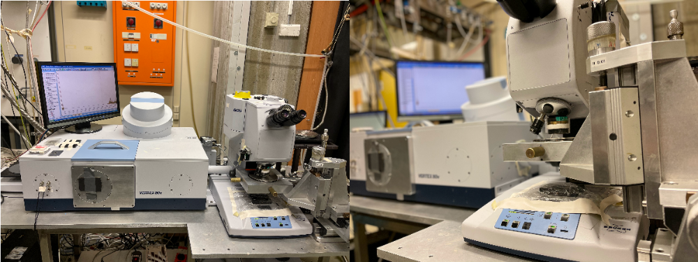
Vertex 80v and IFS 66v/S
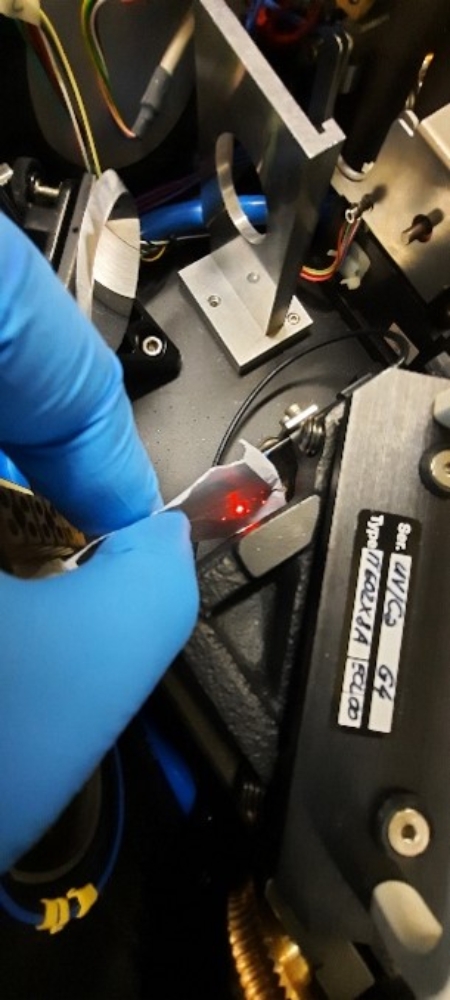
Small samples with dimensions down to the low µm range, for instance, often found in organic materials, are challenging in optical experiments. For this reason, Vertex 80v and IFS 66v/S are equipped with Hyperion IR-microscopes. Here, reflectance and transmittance can be determined with a high spatial resolution and temperature-dependent, inside a LiHe-flow cryostat. For different spectral ranges, a large variety of infrared detectors (Bolometers, DTGS, MCT, Photovoltaic, InSb, Si-diodes) is available. Furthermore, polarization-dependent measurements are possible.
Pressure-dependent optical measurements are realized at the IFS 66v/S with a piston pressure cell (low-temperature low-pressure setup [Link: https://www.pi1.uni-stuttgart.de/research/methods_overview/high-pressure-methods/]) or with a diamond anvil cell at IFS 66v/S and Vertex 80v.
Both spectrometer support time resolved step-scan measurements. For this purpose, at Vertex 80v a Nd:YAG-Laser is attached.
IFS 120v
The Bruker IFS 120v offers ultra-high resolutions up to 0.0016 cm-1. Here, for instance, spectroscopy of molecular vibrations, characterized by a very sharp lineshape, is possible.

Literature:
[some recent papers of institute members]
https://journals.aps.org/prl/abstract/10.1103/PhysRevLett.125.076403
https://journals.aps.org/prb/abstract/10.1103/PhysRevB.101.115118
https://journals.aps.org/prb/abstract/10.1103/PhysRevB.101.161115
[books and review articles]
1. Dressel and G. Grüner, Electrodynamics of Solids: Optical Properties of Electrons in Matter (Cambridge University Press, 2002)
2. B. Tanner, Optical Effects in Solids (Cambridge University Press, 2019)
3. N. Basov, R. D. Averitt, D. van der Marel, M. Dressel, and K. Haule, Rev. Mod. Phys. 83, 471 (2011)